The birth of a Revolution
Few technologies have significantly impacted mass cytometry in the ever-evolving landscape of cellular analysis. This revolutionary technique has transformed our ability to peer into the intricate world of cells, offering unprecedented insights into their complexity and behavior.
The story of mass cytometry begins, like many scientific breakthroughs, with a moment of inspiration. Legend has it that Scott Tanner, a professor at the University of Toronto, having the idea of using the precision of mass spectrometry to analyze cells. This seemingly simple idea would lead to a paradigm shift in single-cell analysis. In 2004, he founded DVS sciences, acquired by Fluidigm in 2014, renamed later Standard BioTools in 2022, reaching 200 millions revenues in 2022.
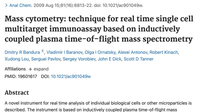
In 2009, Tanner and his team, including Dmitry Bandura, Vladimir Baranov, and Olga Ornatsky, published their groundbreaking paper introducing mass cytometry to the world (Bandura et al., 2009). Their technique combined the cellular analysis capabilities of flow cytometry with the high-resolution mass analysis of inductively coupled plasma mass spectrometry (ICP-MS).
From Flow to Mass: A Quantum Leap
To appreciate the significance of mass cytometry, we must first understand its predecessor, flow cytometry. Developed in the 1960s, flow cytometry has been the workhorse of cellular analysis for decades. It uses fluorescent labels to tag cellular components and then analyzes the fluorescence emitted by individual cells as they flow past lasers.
While revolutionary in its time, traditional flow cytometry has limitations. The overlap between fluorescent spectra restricts the number of cellular features that can be simultaneously analyzed, typically to around 15-20 parameters. This limitation hinders a comprehensive understanding of cellular behavior and complexity. Enter mass cytometry, which shatters this ceiling.
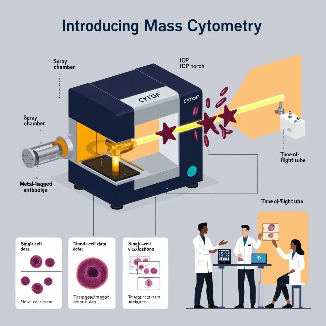
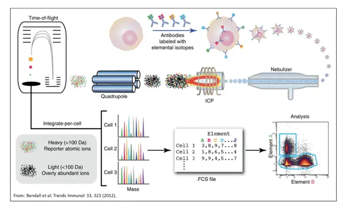
Mass cytometry replaces fluorescent labels with highly pure, stable isotopes of rare earth metals. These metal-tagged antibodies are used to label cellular proteins or other biomolecules. The cells are then vaporized and ionized in a plasma, and the resulting ion clouds are analyzed by time-of-flight mass spectrometry.
The result? A quantum leap in the number of cellular parameters that can be simultaneously measured – up to 50 or more! This dramatic increase in dimensionality allows researchers to capture a much more comprehensive snapshot of cellular states and populations.
The Magic Behind the Curtain
At its core, mass cytometry is a marriage of clever chemistry and sophisticated physics. The process begins with carefully selecting and purifying metal isotopes, each chosen for its unique mass-to-charge ratio. These metals are then conjugated to antibodies using specialized chelating polymers.
Once the cells are labeled, they embark on a journey that would make any amusement park ride seem tame. They’re nebulized into tiny droplets, flash-vaporized at over 7000°C, and turned into a cloud of ions. These ions then race through a flight tube, with heavier ions taking longer to reach the detector than lighter ones.
It’s a process that happens in the blink of an eye – or, more accurately, in about 13 microseconds per cell. Yet, a wealth of information is captured in that brief moment, painting a detailed picture of each cell’s protein expression profile.
Anecdotally, early adopters of mass cytometry often speak of their awe at first seeing the richness of the data. We could describe it as ‘like suddenly being able to see in color after a lifetime of black and white.’ This awe-inspiring moment is a testament to the transformative power of mass cytometry, igniting a sense of inspiration and amazement in those who witness its capabilities.
The Impact and Promise
Mass cytometry has opened up new avenues of research across various fields, from immunology to cancer biology. It has allowed scientists to uncover previously hidden cell subpopulations, track cellular differentiation with unprecedented detail, and gain new insights into complex diseases.
For instance, mass cytometry has revealed the intricate dance of immune cells in response to pathogens, helping unravel our immune system’s mysteries. Cancer research has shed light on tumor heterogeneity, potentially paving the way for more targeted therapies.
Mass cytometry continues to evolve as we stand on the threshold of a new era in cellular analysis. Newer techniques like imaging mass cytometry push the boundaries further, allowing for studying cells within their tissue context.
Mass cytometry has evolved from a beer-inspired idea to a transformative technology. The following chapters explore how this powerful tool reshapes our understanding of biology and medicine. The journey of discovery that began with Tanner and his team continues, promising even more exciting revelations in the years to come. This potential for further discovery and advancement in the field of mass cytometry instills a sense of hope and optimism for the future of cellular analysis.
As for me, well, I stumbled into the world of mass cytometry like a drunk penguin into a black-tie event. There I was, a fresh-faced PhD student, proudly declaring I'd do "pure biology" because computers and I got along like oil and water. Fast forward a few years, and I'm not only writing a book about mass cytometry but I've spawned an algorithm too. Life's funny like that. Even when I tried to escape by adding a medical degree to my academic circus, technology followed me like a clingy ex. I found myself learning from apps and answering digital quizzes, as if the universe was saying, "Nice try, buddy." So here I am, the once computer-phobic biologist, now surfing the wave of high-dimensional data. If my younger self could see me now, he'd probably faint... or at least give his houseplant a very confused look.